C9orf72: what lies within
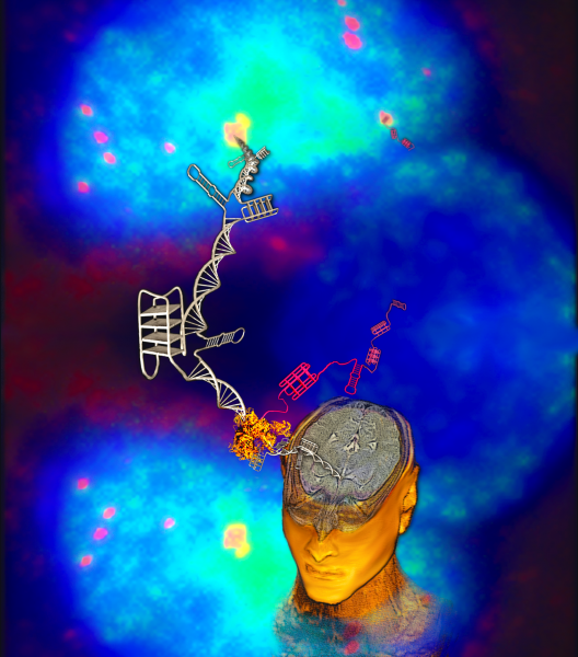
This self portrait was created using an MRI image of the artist's brain and head, which were 3D rendered in the program OsiriX. The DNA (silver) and RNA (pink) were created in Blender. The crystal structure of the RNA polymerase II (orange) was imported from the protein data bank and manipulated using Blender. the image of the neuronal nuclei was obtained via confocal microscopy. The blue is Hoescht dye, which stains for nucelar DNA. The pink dots in the background are RNA that were visualized by Cy5-labeled RNA FISH probes against the repeat RNAs that are transcribed (observed in the foreground). The green is the protein, nucleolin, that was stained with nucleolin-specific primary antibodies and Alexa 488 tagged secondary antibodies. Yellow represents the co-localization of hexanucleotide repeat RNA with nucleolin (Cy5 + Alexa488). All of the images were compiled and edited in the software GIMP.